Magnetic Resonance Imaging (MRI) has revolutionized the field of medical diagnostics with its ability to generate detailed images of the human body rf protection. This non-invasive imaging technique utilizes powerful magnets and radio waves to capture high-resolution images of internal structures, providing valuable insights into various medical conditions.
From diagnosing tumors and evaluating joint injuries to studying brain activity, MRI has become an indispensable tool for healthcare professionals. However, it is crucial to understand both the benefits and limitations of this technology to ensure its optimal utilization and patient safety.
History and Development of MRI
MRI, or magnetic resonance imaging, has a rich history that dates back to the early 1970s. This technology revolutionized medical imaging by providing detailed images of the human body without the need for invasive procedures rf cabin. The development of MRI was a result of advancements in physics, specifically in the field of nuclear magnetic resonance (NMR).
The first MRI machine was built by Raymond Damadian in 1971, and it used a superconducting magnet to generate a strong magnetic field. Over the years, MRI technology has undergone significant advancements, making it more efficient and capable of producing higher resolution images. These advancements include the development of stronger magnets, improved imaging sequences, and the incorporation of advanced computer algorithms for image reconstruction.
MRI has become an indispensable tool in modern medicine, enabling physicians to diagnose and monitor a wide range of diseases and conditions.
How Does MRI Work
Magnetic resonance imaging, commonly referred to as MRI, is a non-invasive medical imaging technique that utilizes strong magnetic fields and radio waves to generate detailed images of the internal structures of the body. This advanced technology has experienced significant advancements over the years, leading to improved image quality, faster scan times, and increased patient comfort.
One notable development is the introduction of higher field strength magnets, which provide clearer and more detailed images. Additionally, the use of parallel imaging techniques has allowed for faster scanning, reducing the time patients spend in the MRI machine.
Future developments in MRI technology aim to further enhance image quality, increase spatial and temporal resolution, and improve the detection and characterization of diseases. These advancements will continue to revolutionize medical imaging, providing clinicians with valuable information to make accurate diagnoses and guide appropriate treatment plans.
Benefits and Limitations of MRI
One of the benefits of utilizing magnetic resonance imaging (MRI) is its ability to provide detailed images of internal structures, allowing for accurate diagnoses and treatment planning. MRI uses a powerful magnetic field and radio waves to generate images of the body.
This non-invasive imaging technique is particularly advantageous in the evaluation of soft tissues, such as the brain, spinal cord, and joints. MRI can detect abnormalities that may not be visible with other imaging modalities, such as X-ray or CT scan.
Additionally, MRI does not involve the use of ionizing radiation, making it a safer option for patients, especially for repeated imaging. However, there are some limitations to MRI. It can be expensive, time-consuming, and requires patients to remain still for extended periods.
Furthermore, certain metallic implants or devices may be incompatible with MRI, limiting its use in some cases.
Common Applications of MRI
MRI is commonly used in the medical field for a variety of applications. This includes the evaluation of brain and spinal cord disorders, detection of tumors, assessment of joint injuries, and monitoring the progression of certain conditions.
In terms of clinical uses, MRI plays a crucial role in diagnosing and understanding various diseases and conditions. It provides detailed images that allow physicians to accurately assess the extent and location of abnormalities. This aids in treatment planning and monitoring.
Additionally, MRI has significant research applications. It is used to study the structure and function of the brain, investigate the effects of different interventions or treatments, and explore the underlying mechanisms of various diseases.
The non-invasive nature of MRI, coupled with its ability to capture detailed anatomical and physiological information, makes it an indispensable tool in both clinical and research settings.
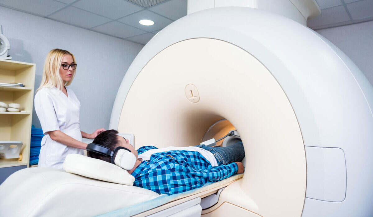
Preparing for an MRI Scan
When preparing for an MRI scan, it is important for patients to remove any metal objects or jewelry that could interfere with the imaging process. MRI machines use powerful magnets to create detailed images of the body’s internal structures. Metal objects can be attracted to the magnet, causing potential risks such as movement artifacts or even injury.
To ensure a successful and safe MRI scan, patients should follow specific preparing techniques. These include removing all metal objects, such as jewelry, piercings, watches, and clothing with metal fasteners. Patients should also inform the medical staff about any implants or devices in their body that may contain metal, such as pacemakers or metal plates.
Conclusion
In conclusion, MRI has revolutionized medical imaging by providing detailed and precise images of the internal structures of the human body. Its non-invasive nature and ability to capture images in different planes make it a valuable tool in diagnosing various conditions and monitoring treatment effectiveness.
Despite its numerous benefits, MRI has limitations such as high cost and limited availability in certain regions. Nonetheless, ongoing advancements in technology continue to enhance the capabilities and accessibility of this imaging modality.


